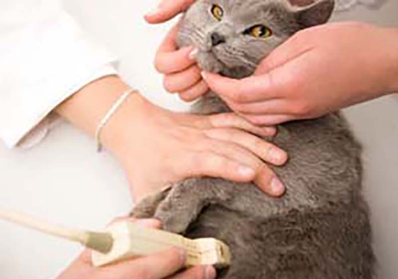
Ultrasound is a non-invasive, state-of-the-art technology that uses sound waves to painlessly examine internal organs, muscles, and vessels. Ultrasound allows deep, three-dimensional visualization of a pet’s internal structures, and can be used in conjunction with x-rays to assess organ function, detect tumors or fluid, determine pregnancy, and more. Ultrasound reveals important information about internal processes including the circulatory, skeletal, and gastrointestinal systems, helping identify disease, blockages and internal injury.
The doctors at LAVet Hospital have obtained extensive additional training on abdominal ultrasonography in dogs and cats. An ultrasound works by broadcasting high-frequency sound waves that reflect off your pet’s internal structures. A small probe held against the skin collects the returning signals to create an image of the internal body, most commonly used to examine abdominal organs like the stomach, kidneys, liver, spleen and gallbladder.
At LAVet Hospital, we routinely use ultrasound to diagnose bladder and kidney stones, intestinal blockages, discover abdominal masses, and to aid in the assessment of organ function.
The dedicated doctors and staff at LAVet Hospital are here when you need us to diagnose your pet’s condition. We are conveniently located to Thibodaux, Houma, Raceland, Lockport, Chackbay, and other neighboring communities.